
Written by Dr. Muhammad Azfar bin Noordin

Dr. Muhammad Azfar bin Noordin
Senior Lecturer
Faculty of Mechanical Engineering
Universiti Teknologi Malaysia
Treating critical bone defects remains one of the greatest challenges in modern orthopedic medicine. Every year, millions of patients require bone grafts to repair fractures caused by trauma, disease, or congenital abnormalities. Although conventional metallic implants provide the structural support needed during healing, they often remain permanently in the body and may require a second surgery to remove after recovery. These additional procedures increase healthcare costs, prolong patient recovery, and expose patients to unnecessary surgical risks. Consequently, researchers worldwide are actively pursuing biodegradable metallic implants that can safely support healing and then gradually disappear once their role is complete.
Among the various biodegradable metals under investigation, iron (Fe) has emerged as a particularly promising candidate because of its excellent mechanical strength and compatibility with human bone. Unlike magnesium, which degrades too rapidly, or zinc, which may not always provide sufficient strength, iron offers a better balance between mechanical performance and structural stability. However, its naturally slow degradation remains a major limitation.
Developing an iron scaffold that degrades at an appropriate rate while maintaining sufficient strength throughout healing has become a key challenge in bone tissue engineering. Addressing this challenge requires a deeper understanding of how implant design and physiological conditions influence degradation inside the human body.
Dr Muhammad Azfar Noordin, who leads a team of researchers in the Department of Applied Mechanics and Design, Faculty of Mechanical Engineering, Universiti Teknologi Malaysia (UTM), has taken an important step toward advancing biodegradable bone implant development through a more physiologically relevant experimental approach. Rather than relying solely on conventional laboratory immersion tests, the research integrates a reconstructed cancellous bone structure with a dynamic simulated body fluid (SBF) system to more closely replicate the conditions implants experience in the human body after implantation, as illustrated in Figure 1.
By recreating the dynamic flow of body fluids through the porous bone environment, this innovative testing platform provides a more realistic assessment of scaffold degradation and mechanical performance. The findings offer valuable insights into how biodegradable porous iron implants interact with their surrounding physiological environment, bringing researchers closer to designing safer, more reliable, and clinically relevant bone scaffolds for future orthopedic applications.
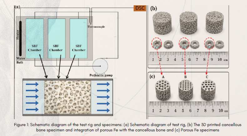
Most laboratory studies evaluate biodegradable implants by immersing them in simulated body fluid under either static or simple flowing conditions. While these approaches provide useful baseline information, they do not accurately represent the complex environment found inside cancellous bone. Human cancellous bone contains an interconnected porous network through which bone marrow continuously circulates, transporting nutrients, oxygen, and metabolic waste while generating fluid-induced shear stress that influences both bone remodeling and implant degradation. Replicating these physiological conditions is essential for producing more realistic predictions of implant performance.
To recreate this environment, the research team harvested bovine cancellous bone and reconstructed its intricate internal architecture using high-resolution micro-computed tomography (micro-CT). The researchers then reproduced the three-dimensional bone model through stereolithography (SLA) additive manufacturing, enabling them to integrate porous iron scaffolds within an anatomically representative cancellous bone structure. Simulated body fluid was circulated continuously through the system at a controlled flow rate, reproducing the movement of bone marrow within living bone and exposing the scaffolds to realistic mechanical and chemical conditions
Researchers fabricated three porous iron scaffold designs with approximately 80% porosity using precision computer numerical control (CNC) machining. Although all specimens possessed similar overall porosity, each employed a different pore configuration and surface area. This allowed the researchers to isolate the influence of pore architecture on degradation behaviour while keeping other important variables, including porosity, material composition, temperature, and flow conditions, constant throughout the investigation.
The scaffolds were immersed under dynamic conditions for 7, 14, and 28 days before undergoing detailed microstructural, chemical, and mechanical characterization.
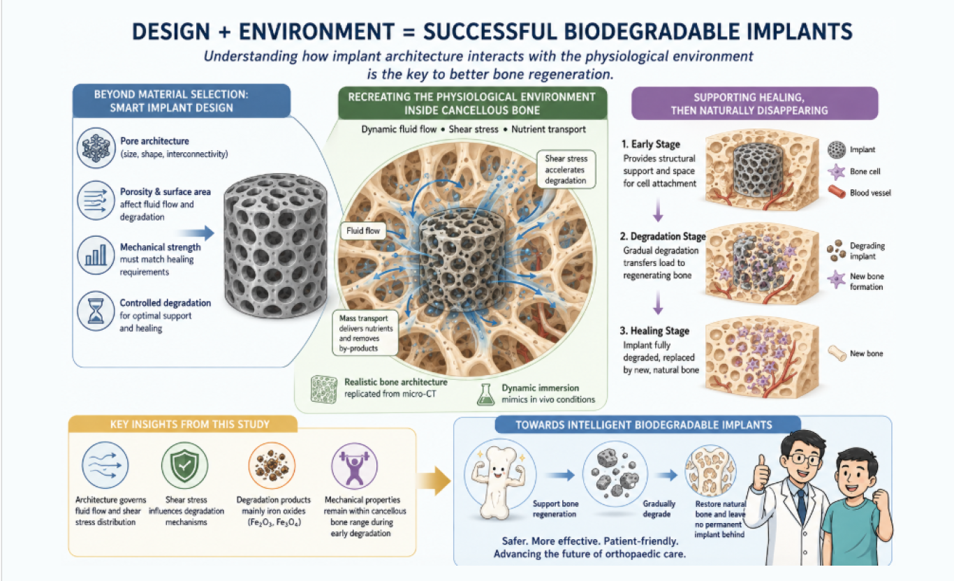
The study shows that scaffold degradation depends not only on material composition but also on pore architecture and fluid flow. Surprisingly, the scaffold with the lowest exposed surface area showed the highest degradation rate, indicating that pore configuration and fluid-induced shear stress can be more important than surface area alone.
Microscopic and chemical analyses revealed iron oxides, mainly Fe₂O₃ and Fe₃O₄, as the dominant degradation products. Regions exposed to higher shear stress also experienced greater material loss, suggesting that scaffold geometry can be designed to control degradation behaviour.
Although Young’s modulus and yield strength decreased by approximately 65–78% over 28 days, the degraded scaffolds retained mechanical properties comparable to natural cancellous bone. This suggests they can continue supporting bone healing while gradually transferring load back to the regenerated tissue.
The research also introduces a more realistic dynamic testing method for biodegradable implants. By combining cancellous bone-like structures with fluid-flow conditions, the approach can improve predictions of implant performance and support the development of personalized biodegradable scaffolds for orthopedic, craniofacial, and dental applications.
